- Cell Wall
- The tasks of the complex bacterial cellwall are to protect the protoplasts from
external noxae, to withstand and maintain the osmotic pressure gradient between
the cell interior and the extracellular environment (with internal pressures
as high as 500–2000 kPa), to give the cell its outer form and to facilitate
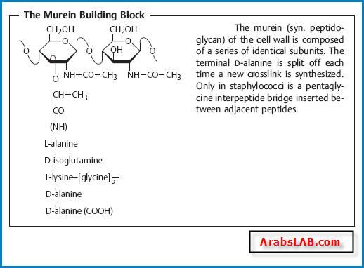
communication with its surroundings. - Murein (syn. peptidoglycan).
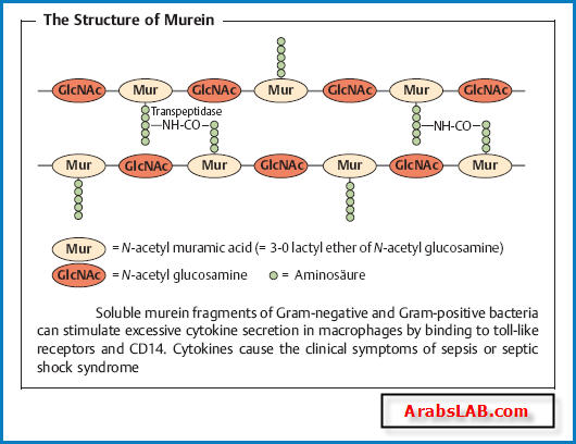
The most important structural element of
the wall is murein, a netlike polymer material surrounding the entire cell
(sacculus). It is made up of polysaccharide chains crosslinked by peptides.
the wall is murein, a netlike polymer material surrounding the entire cell
(sacculus). It is made up of polysaccharide chains crosslinked by peptides.
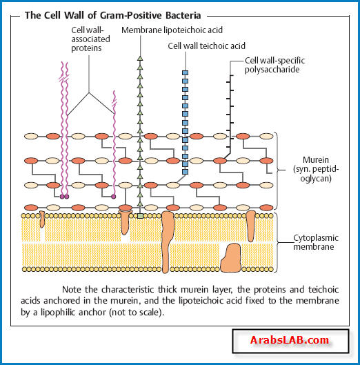
The cell wall of Gram-positive bacteria.
The murein sacculus may
consist of as many as 40 layers (15–80 nm thick) and account for as much as
30% of the dry mass of the cell wall. The membrane lipoteichoic acids are
anchored in the cytoplasmic membrane, whereas the cell wall teichoic acids
are covalently coupled to the murein. The physiological role of the teichoic
acids is not known in detail; possibly they regulate the activity of the autolysins
that steer growth and transverse fission processes in the cell. Within
the macroorganism, teichoic acids can activate the alternative complement
pathway and stimulate macrophages to secrete cytokines. Examples of cell
wall-associated proteins are protein A, the clumping factor, and the fibronectin-
binding protein of Staphylococcus aureus or theMprotein of Streptococcus
pyogenes. Cell wall anchor regions in these proteins extending far beyond the
murein are bound covalently to its peptide components. Cell wall-associated
proteins frequently function as pathogenicity determinants (specific adherence;
phagocyte protection).
The cellwall of Gram-negative bacteria.
Here, the murein is only about 2 nm
thick and contributes up to 10% of the dry cell wall mass . The outer
membrane is the salient structural element. It contains numerous proteins
(50% by mass) as well as the medically critical lipopolysaccharide.
thick and contributes up to 10% of the dry cell wall mass . The outer
membrane is the salient structural element. It contains numerous proteins
(50% by mass) as well as the medically critical lipopolysaccharide.
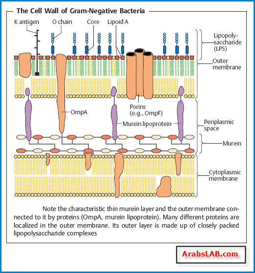
- Outer membrane proteins.
— OmpA (outer membrane protein A) and the murein lipoprotein form a
bond between outer membrane and murein.
— Porins, proteins that form pores in the outer membrane, allow passage of
hydrophilic, low-molecular-weight substances into the periplasmic space.
— Outer membrane-associated proteins constitute specific structures that
enable bacteria to attach to host cell receptors.
— A number of Omps are transport proteins. Examples include the LamB
proteins for maltose transport and FepA for transport of the siderophore
ferric (Fe3+) enterochelin in E. coli .
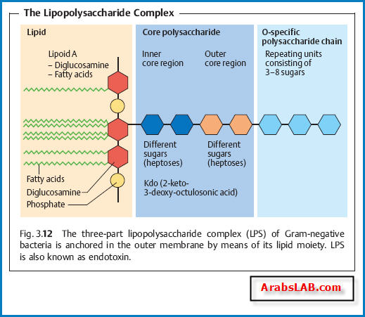
- Lipopolysaccharide (LPS).
This molecular complex, also known as endotoxin,
is comprised of the lipoid A, the core polysaccharide, and the O-specific
polysaccharide chain .
is comprised of the lipoid A, the core polysaccharide, and the O-specific
polysaccharide chain .
- Lipoid A
is responsible for the toxic effect. As a free substance, or bound up
in the LPScomplex, it stimulates—by binding together with the LPS binding
protein (LBP) to the CD14 receptor of macrophages—the formation and
secretion of cytokines that determine clinical endotoxin symptomatology.
Interleukin 1 (IL-1) and tumor necrosis factor (TNF) induce an increased
synthesis of prostaglandin E2 in the hypothalamus, thus setting the “thermostat”
in the temperature control center higher, resulting in fever. Other
direct and indirect endotoxin effects include granulopoiesis stimulation,
aggregation and degeneration of thrombocytes, intravasal coagulation due
to factor VII activation, a drop in blood pressure, and cachexia. LPS can also
activate the alternative complement pathway. Release of large amounts of
endotoxin can lead to septic (endotoxic) shock. Endotoxin is not inactivated
byvapor sterilization. Therefore, the parent materials usedinproduction of
parenteral pharmaceuticals must be free of endotoxins (pyrogens).
- The O-specific polysaccharide chain
is the so-called O antigen, the fine
chemical structure of which results in a large number of antigenic variants
useful in bacterial typing (e.g., detailed differentiation of salmonella
types) .
chemical structure of which results in a large number of antigenic variants
useful in bacterial typing (e.g., detailed differentiation of salmonella
types) .
- L-forms (L = Lister Institute).
The L-forms are bacteria with murein defects,
e.g., resulting from the effects of betalactam antibiotics. L-forms are highly
unstable when subjected to osmotic influences. They are totally resistant to
betalactams, which block the biosynthesis of murein. The clinical significance
of the L-forms is not clear. They may revert to the normal bacterial form when
betalactam therapy is discontinued, resulting in a relapse.
- Capsule
Many pathogenic bacteria make use of extracellular enzymes to synthesize a
polymer that forms a layer around the cell: the capsule. The capsule protects
bacterial cells from phagocytosis. The capsule of most bacteria consists of a
polysaccharide. The bacteria of a single species can be classified in different
capsular serovars (or serotypes) based on the fine chemical structure of this
polysaccharide.
bacterial cells from phagocytosis. The capsule of most bacteria consists of a
polysaccharide. The bacteria of a single species can be classified in different
capsular serovars (or serotypes) based on the fine chemical structure of this
polysaccharide.
- Flagella
Flagella give bacteria the ability to move about actively. The flagella (singular
flagellum) are made up of a class of linear proteins called flagellins. Flagel
lated bacteria are described as monotrichous, lophotrichous, or peritrichous,
depending on how the flagella are arranged . The basal
body traverses the cell wall and cytoplasmic membrane to anchor the flagellum
and enables it to whirl about its axis like a propeller.
In Enterobacteriaceae, the flagellar antigens are called H antigens. Together
with the O antigens, they are used to classify bacteria in serovars.
- Attachment Pili (Fimbriae), Conjugation Pili
Many Gram-negative bacteria possess thin microfibrils made of proteins (0.1–
1.5 nm thick, 4–8 nm long), the attachment pili. They are anchored in the
outer membrane of the cell wall and extend radially from the surface. Using
these structures, bacteria are capable of specific attachment to host cell receptors
(ligand—receptor, key—keyhole).
The conjugation pili (syn. sex pili) in Gram-negative bacteria are required
for the process of conjugation and thus for transfer of conjugative plasmids.
- Biofilm
A bacterial biofilm is a structured community of bacterial cells embedded in a
self-produced polymer matrix and attached to either an inert surface or living
tissue. Such films can develop considerable thickness (mm). The bacteria located
deep within such a biofilm structure are effectively isolated from immune
system cells, antibodies, and antibiotics. The polymers they secrete are
frequently glycosides, from which the term glycocalyx (glycoside cup) for the
matrix is derived.
- Bacterial Spores
Bacterial spores (endospores) are purely dormant life forms. Their development
frombacterial cells in a “vegetative” state does not involve assimilation
of additional external nutrients. They are spherical to oval in shape and are
characterized by a thick spore wall and a high level of resistance to chemical
and physical noxae. Among human pathogen bacteria, only the genera Clostridium
and Bacillus produce spores. The heat resistance of these spores is
their most important quality from a medical point of view, since heat ster
ilization procedures require very high temperatures to kill them effectively.
Potential contributing factors to spore heat resistance include their thickwall
structures, the dehydration of the spore, and crosslinking of the proteins by
the calcium salt of pyridine-2,6-dicarboxylic acid, both of which render protein
denaturing difficult. When a spore’s milieu once again provides favorable
conditions (nutrient medium, temperature, osmotic pressure, etc.) it returns
to the vegetative state in which spore-forming bacteria can reproduce.

0 التعليقات:
Post a Comment